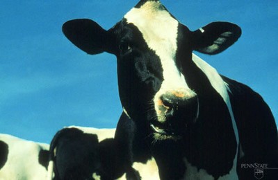
New Mexico is classified FREE of BRUCELLOSIS
To prevent introduction of the disease brucellosis into New Mexico and to comply with United States Department of Agriculture regulations pertaining to Brucellosis, animals from non-free status states must meet the following requirements.
At this time the only Class A non-free state is Montana.
Breeding age animals sold within the state of New Mexico are tested at sale barns as a surveillance program.
Beef Cattle imported from non free states must meet the following requirements:
- All breeding age animals 18 months of age and older negative test required.
- If proof of prior calfhood vaccination, a negative brucellosis test is required if over 24 months of age.
- Dairy Cattle imported from non free states, a negative test is required of all breeding animals if over 18 months of age.
Dairy Goats imported from non free states must have a negative brucellosis test, unless originating from a certified Brucellosis free herd.
Calfhood Brucellosis vaccination with a federally approved vaccine is recommended to allow flexible interstate movement. Regulations concerning vaccination requirements vary with each state.
- Beef heifers should be vaccinated between 4 to 12 months of age.
- Dairy heifers should be vaccinated between 4 to 10 months of age.
Equine infectious anemia (EIA) is a viral disease caused by a lentivirus, a member of the family known as retroviruses. Retroviruses incorporate their genetic material (DNA) into the DNA of host cells, and generally infect the host for life. In the case of EIA the viral DNA is combined with the DNA of the host's white blood cells. The disease has been referred to by several common names, such as swamp fever, mountain fever, and slow fever.
The virus is transmitted to horses mechanically on the mouthparts of biting flies such as horse flies and deer flies, thus transmission is more common in the spring, summer and early fall. It can also be transmitted by hypodermic needles, and from an infected mare to foal in utero. The disease is found worldwide, more commonly in humid, swampy or boggy areas. The incubation period can vary from a week to a few months.
Clinical signs are often nonspecific or inapparent. In the acute phase of the infection they can include fever, weakness, anemia, jaundice, hemorrhages on the gums and blood-stained feces. Horses may die during the acute phase, but more likely they can become chronic, often inapparent carriers. Some carriers will develop episodic symptoms when virus is released into the bloodstream, especially when stressed by other illnesses, hard work severe stress. During these periods biting flies are more likely to pick up and transmit the virus from an infected animal.
The disease can be detected by enzyme-linked immunosorbent assay (ELISA) and agar gel immunodiffusion (AGID, aka Coggins) tests. There is no vaccine available for the disease, nor is there any curative treatment. Any horse six months of age or older being brought into New Mexico is required to have a negative AGID or ELISA test for EIA within 12 months of entry, except for nursing foals accompanying EIA test-negative dams.
Statutory Rule for EIA Testing
77-3-14.1. AGID tests required.
The board shall adopt rules prohibiting the driving or transporting into this state of any horses or other equidae that have not tested negative to the AGID, or Coggins, test or a United States department of agriculture-approved equivalent test for equine infectious anemia within twelve months prior to the date of entry, the evidence of which test result shall be shown on a health certificate; excepting from regulation only those foals accompanied in shipment by a negative-tested dam, those horses or other equidae consigned directly to slaughter.
NMAC for Equine Imports
21.32.4.13 IMPORT REQUIREMENTS FOR EQUIDAE:
A. All equidae, which includes horses, mules and asses, entering New Mexico must be accompanied by an official health certificate attesting the equidae in the shipment are free from symptoms of infectious or contagious disease.
B. All equidae entering the state of New Mexico must be tested and negative, within 12 months prior to entry, for equine infectious anemia (EIA) using the agar gel immunodiffusion (AGID) test, also known as the "Coggins" test or the competitive enzyme-linked immunosorbent assay (CELISA) test or other USDA licensed test approved by the board. The date of the test, the laboratory and the results must be shown on the required health certificate. Individual identification and/or description of the animal(s) must also be provided on the health certificate.
C. Foals, nursing and accompanied in shipment by a negative (EIA) tested dam and equidae consigned directly to slaughter in New Mexico are not required to be tested for EIA. If the dam does not accompany the foal in shipment, the foal must be tested negative prior to entry.
D. All testing for EIA must be performed at laboratories approved by USDA for such testing. All samples must be collected by an accredited veterinarian or full-time state or federal regulatory personnel.
E. The state veterinarian may grant a special permit to enter the state of New Mexico for equidae that have a test pending. This permit must be requested and granted prior to entry.
[3/1/99; 21.32.4.13 NMAC - Rn, 21 NMAC 32.4.13, 12/31/2007]
Equine viral arteritis (EVA) is a contagious viral disease (equine arteritis virus) of horses worldwide. Limited date suggests the virus may also infect alpacas and llamas. Disease transmission may occur directly by aerosol (principal route) , congenital, and venereal routes (to include artificial insemination) and indirectly by contact with fomites, urine, feces, and vaginal secretions). The majority of acute EVA infections are inapparent (subclinical). Clinical signs of disease are variable, but may include fever, depression, anorexia, dependent edema (limbs, scrotum, prepuce, mammary tissue, lower abdomen), abortion, conjunctivitis, rash, and rhinitis. EVA is of special economic concern because it can result in the establishment of the carrier state in stallions, abortion in pregnant mares, and illness and death in young foals. Vaccination appears to prevent uninfected stallions from becoming long term carriers. EVA is a disease that is reportable to the State and Federal Animal Health Officials.
Additional Information:
Authorities:
NM Statute and Rule
NMAC 21.30.7. (1-21)
USDA APHIS Equine Viral Arteritis Uniform Method and Rules 91-55-075
The New Mexico Equine Viral Arteritis Rule (NMAC 21.30.7.19) specifies that:
- Issue of EVA vaccine is to federally accredited New Mexico-licensed veterinarians following written request through the state veterinarian.
- Testing of stallions for antibodies in blood or evidence of EAV in semen is to be done at an approved veterinary laboratory.
- Before an initial EVA vaccination, stallions must be confirmed EVA test negative to a blood sample collected by an accredited veterinarian.
- Intact colts between six (6) and twelve (12) months of age must be confirmed EVA Test sero-negative before vaccination.
- Initial EVA vaccination of stallions be done by an accredited veterinarian within ten (10) days of the pre-vaccination sample collection date.
- Following the initial EVA vaccination, an equid shall not have direct exposure to an EVA affected animal or pregnant mare for twenty-eight (28) days after vaccination.
- Following EVA vaccination, a vaccinated stallion shall not be used for breeding or artificial insemination within twenty-eight (28) days after vaccination. A vaccinated mare shall not be bred within twenty-one (21) days of vaccination.
See the New Mexico EVA Rule for other requirements.
Ordering Process for Equine Viral Arteritis Vaccine
In New Mexico, Equine Viral Arteritis Vaccine is registered as a restricted use vaccine. Firms authorized to distribute the product must receive State Veterinarian authorization before distribution to the accredited veterinarian. The New Mexico Equine Viral Arteritis Rule (NMAC 2.30.7.19) specifies that federally accredited New Mexico-licensed veterinarians submit a written request to the State Veterinarian to obtain Equine Viral Arteritis (EVA) Vaccine for use.
The process for federally accredited New Mexico-licensed veterinarian to request and obtain Equine Viral Arteritis Vaccine is as follows:
- Complete the form entitled, Request to Obtain Equine Viral Arteritis Vaccine, Arvac.
Note: NMLB will only process legible and complete forms
and will return illegible and incomplete forms to the requestor.
- Forward the Request to Obtain Equine Viral Arteritis Vaccine, Arvac by email directly to the New Mexico State Veterinarian by email at: statevet@nmlbonline.com
EVA Vaccine Request Form
Request to Obtain Equine Viral Arteritis Vaccine, Arvac
Upon receipt of the Request to Obtain Equine Viral Arteritis Vaccine, Arvac, the State Veterinarian, or designee, will:
- Review and log in the vaccine request.
- Obtain any additional necessary information.
- Approve the request and complete The State Veterinarian Authorization / Distribution Permit section.
- Email the approved request directly to the authorized distributor of the vaccine (MWI)
Requirement for Certification of Vaccination
The accredited veterinarian submit an official Certificate of EVA Vaccination (prescribed by the State Veterinarian) to the State Veterinarian within seven (7) days of the vaccination date. [NMAC 21.30.7.19(E)]
- For initial EVA vaccination, the original EVA Test laboratory report is to accompany the Certificate of EVA Vaccination
Note: Contact the New Mexico Horse Breeders' Association for Stallion Registration procedures and specific EVA vaccination certification requirements.
What is Johne's Disease?
Johne's Disease is a chronic debilitating disease of cattle caused by Mycobacterium avium paratuberculosis, a bacteria related to M. bovis, the cause of bovine tuberculosis. It is spread by ingestion of feces containing the organisms, with transmission most likely in pre-weaning calves born in contaminated calving quarters and nursing from contaminated teats. Less commonly the disease is spread calf-to-calf and to calves in utero.
The organism undergoes a prolonged incubation period before clinical signs are evident. The infection causes a marked thickening of the intestinal wall and decreased ability to absorb nutrients, which leads to signs including chronic diarrhea, weight loss and decreased milk production. Infected individuals may shed the organisms intermittently or constantly. Some individuals which shed very large numbers of the organism have been termed super shedders. Infected cows can be detected by fecal culture or milk ELISA testing. Veterinary Diagnostic Services, operated by the NM Dept. of Agriculture and located in Albuquerque, is equipped to run milk ELISA testing.
Because the progression of signs is insidious producers often fail to recognize the effects on their herd, or discount the effects as not being economically important enough to try to control.
Control measures include:
- Keep clinical cases and JD test-positive cows out of the maternity pen.
- Clean the maternity pen often.
- Get the calf out of the maternity pen in less than an hour after birth.
- Feed each calf 4 quarts (3 for Jerseys) of clean colostrum before the calf is 6 hours old.
- Teats should be prepped before colostrum collection to limit manure contamination.
- Colostrum should be fed from one test-negative cow to one calf. Identification of the cow donating the colostrum to the calf should be recorded in the calf's record.
- If colostrum is pooled, it should only be from test-negative cows, or pasteurized.
- After colostrum, feed only pasteurized milk.
- Milk replacer or on-farm pasteurized milk is acceptable, if the on-farm pasteurizer is regularly maintained.
- On-farm pasteurizers should be checked monthly for significant decrease in milk total bacterial counts between pre- and post-pasteurization.
- Rear calves well away from adult cattle insuring no contact with manure or contamination of water or feed with manure.
June 2016
The SFCP Standards Have Been Revised
The June 2013 Scrapie Free Flock Certification Program (SFCP) standards have been updated and are available on the APHIS SFCP Web page. The May 2016 SFCP standards are now in effect.
A brief summary of the major updates to the program are also available on the SFCP Web page.
The basic structure of the program has not changed. There are still two categories in the SFCP: the Export Category (with Export Monitored flocks and Export Certified flocks), and the Select Category (Select Monitored flocks). The updates address/clarify:
- Sampling requirements, advancement, and genotyping lambs/kids in genetically resistant flocks;
- Veterinary inspection of cull animals;
- Imported embryos/oocytes;
- Animals originating from Inconsistent States;
- Special circumstances involving "Lost to Inventory" and "Found Dead" animals; and
- Reporting requirements for the use of milk/colostrum from a lower status flock.
About Scrapie
Scrapie is a chronic progressive fatal disease of the central nervous system in sheep and goats. It is one of several diseases known as transmissable spongiform encephalopathies, or TSE's. Other members of this group of diseases are bovine spongiform encephalopathy, (BSE or "Mad Cow" disease), chronic wasting disease in deer, and Jacob-Cruetzfeld disease in man. These diseases are caused by abnormal prion proteins in the brain. There is a genetic difference among sheep for the tendency to form these abnormal proteins, and sheep can be genotyped for susceptibility. The susceptibility to the disease is passed on to lambs with certain genes from parents that carry the genes for susceptibility.
The disease is slow to develop and progress. Early signs include restlessness, weight loss, itching and rubbing, wool pulling, abnormal gait, and tremors. Eventually the signs progress to the point that the affected animal goes down and is unable to stand, and the animal eventually dies. The course of the disease may be as long as 2-3 years after clinical signs are first noted.
Initially a diagnosis could only be made by microscopic examination of brain tissue from a deceased animal, but in recent years microscopic exam of lymphoid tissue from the third eyelid or rectum has been shown as a way to diagnose the disease in the living animal. There is no treatment for the disease, nor is there any vaccine for prevention.
Frequently Asked Questions
Click the link below for a document answering a number of frequent questions about scrapie.


